How To Calculate Pixel Size Mri
At a map scale of 1 to 75000 one pixel refers to a. Pixel sizes range in clinical MRI from mm eg 1 3 1 mm2 to sub-mm.
Their diameters are generally measured in micrometers microns.
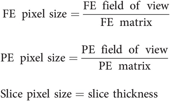
How to calculate pixel size mri. Calculate the pixel size for a fast spin echo sequence with the following parameters. If you have segmented a tumor you can multiply the number of segmented voxels by the voxel volume dx dy dz which can be obtained e. FOVfNf field of view in the frequency encoding direction divided by the number of steps in the frequency encoding direction Pixel area for 2D sequences Pixel size in the read or frequency direction times the pixel size in the phase direction.
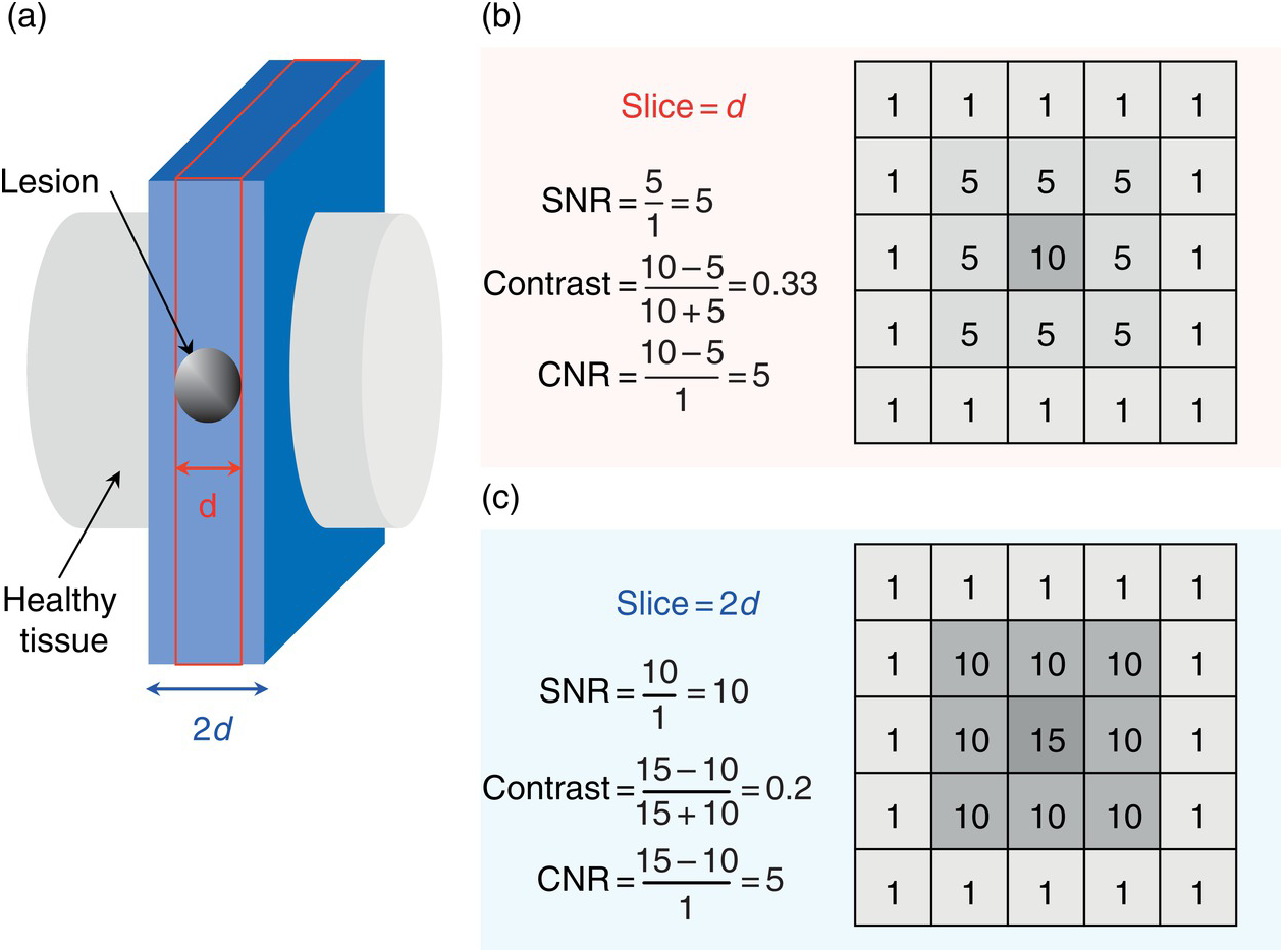
Higher resolution means more image detail for example when two structures 1 mm apart are distinguishable in an image this picture has a higher resolution than an image where they are not to distinguish. Basic resolution and phase resolution. Its dimensions are given by the pixel together with the thickness of the slice the measurement along the third axis.
Pixel s do not have a fixed size. Hence increasing the field of view in either direction increases the size of the voxels and decreases the resolution. Decreasing the field of view improves the resolution.
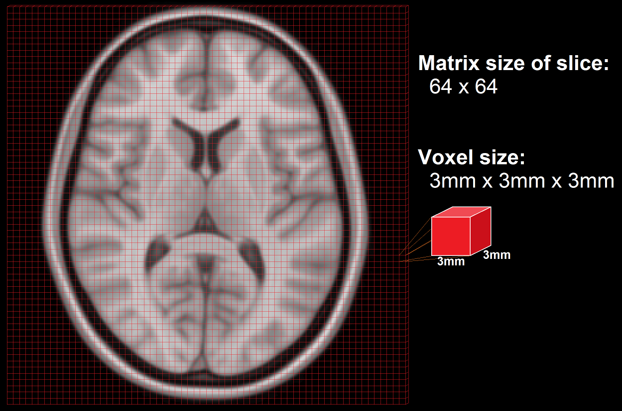
A voxel is the volume element defined in 3D space. FOV 30cm TR 600 TE 10 320 x 320 matrix 4 NEX 3 mm slice thickness 3 ETL. FOVfNf The field of view in the frequency encoding direction divided by the number of steps in the.
Although the field-of-view and pixel size may vary in each of the cardinal directions for clarity of explanation we will consider only the symmetric 2-dimensional. The matrix size is typically 128x 256x or 512x. Slice thicknesses in clinical MRI.
Digitized it has 157 times 79 pixels those are 12403 pixels. It has dimensions given along two axes in millimeters dictating in-plane spatial resolution. Pixel size can be calculated by dividing the field of view by the matrix size egFOV 320 Matrix 320x320 Pixel size 3203201mm.
Pixel size is typically between 05 and 15 mm. The pixel size is equal to the field of view divided by the matrix size. Dividing the field of view by the matrix size gives you the in-plane voxel size.
Now i want to compare these measurements statistically. The FOV is typically divided into several hundred picture elements pixels each approximately 1 mm² in size. The image resolution is the level of detail of an image and a measurement of image quality.
Multiply phase pixel size by frequency pixel size pixel area squared Frequency dimension of a pixel FOV frequency matrix. More data points in an MR image with same FOV will decrease the pixel size. Pixel sizes range in clinical MRI from millimeters eg 11 mm 2 to submillimeters.
Field-of-view FOV refers the distance in cm or mm over which an MR image is acquired or displayed. Pixel size can be calculated by dividing the field of view by the matrix size egFOV 320 Matrix 320x320 Pixel size 3203201mm. Its dimensions are given by the pixel together with the thickness of the slice the measurement along the third axis.
Pixel size FOVfrequencyphase value Example. Encoding direction equals the pixel size in the phase encoding direction. There are two resolution parameters used in MRI for the production of a two dimensional.
There are two resolution parameters used in MRI for the production of a two dimensional image ie. We can calculate the size of our pixel by taking the field of view FOV and dividing it by the frequencyphase value. Pixel size in the read or frequency direction.
Although the pixel is not a unit of measurement itself pixel s are often used to measure the resolution or sharpness of images. Voxel volume for 3D sequences. 094 mm x 094 mm FOV Matrix pixel size FOV phase matrix phase dimension pixel.
Given along two axes in mm dictating in-plane spatial resolution. Clicking Area multiplies the number of pixels in width and length. Frequency256 Phase192 FOV200 200256 Frequency direction 200192Phase direction This would create a pixel with 78mm x 104mm.
Multiply phase pixel size x frequency pixel size Pixel area answer squared 2. A map detail scanned with 200 dpi has an original length of 2 cm and width of 1 cm. G from the DICOM tags Pixel.
To determine the pixel size in the read or frequency direction use this formula. A voxel is the volume element defined in 3D space. I have been used image j program to calculate pixel intensity values in different DICOM CT images.
Pixel size is dependent on both the field of view and the image matrix.

Mri Resolution And Image Quality How To Manipulate Mri Scan Parameters
%20and%20SNR%20relation%20in%20MRI.jpg)
Signal To Noise Ratio Snr In Mri Factors Affecting Snr Calculating Snr Mri

Mri Resolution And Image Quality How To Manipulate Mri Scan Parameters

Nipype Beginner S Guide All You Need To Know To Become An Expert In Nipype

Mri Resolution And Image Quality How To Manipulate Mri Scan Parameters

Mr Image Quality Frcr Physics Notes

Mr Image Quality Frcr Physics Notes

Mr Image Quality Frcr Physics Notes

The Devil S In The Detail Pixels Matrices And Slices Chapter 5 Mri From Picture To Proton

Mri Resolution And Image Quality How To Manipulate Mri Scan Parameters

Mri Resolution And Image Quality How To Manipulate Mri Scan Parameters

What You Set Is What You Get Basic Image Optimization Chapter 6 Mri From Picture To Proton
Post a Comment for "How To Calculate Pixel Size Mri"